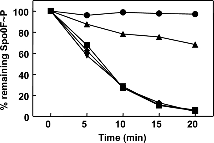
Fig 6.

Activities of the RapAC2 and RapAC3 hybrid proteins. Time courses of dephosphorylation of Spo0F∼P by RapAC2 and RapAC3 were carried out in vitro as described in Materials and Methods. Proteins were used at a 1.5 μM concentration. The RapAC2 hybrid protein was also tested in the presence of PhrC (3 μM). The samples were run on a 15% SDS-PAGE gel, exposed to a PhosphorImager screen, and quantified by the ImageQuant software program. Symbols: ●, Spo0F∼P alone; ■, Spo0F∼P and RapA wt; ▼, Spo0F∼P and RapAC2; ◆, Spo0F∼P and RapAC3; ▲, Spo0F∼P, RapAC2, and PhrC.
