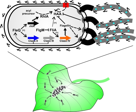
Fig 1.
P. syringae swarming with surfactant and flagella. A population of P. syringae cells exhibiting active swarming motility on a bean leaf surface are depicted (bacteria are not drawn to scale). Bacterial motility is indicated with curved arrows showing the cells converging on nutrient-rich sites on the plant surface where bacterial aggregates form. An enlarged bacterial cell is shown with a small tuft of polar flagella (extending off the page; P. syringae generally produces 2 to 4 polar flagella). HAA is synthesized by the activity of RhlA from acylated precursors. HAA is indicated as the linked squiggles reflective of its dual branched structure (not drawn to scale). HAA is exported from the cell by an uncharacterized export machinery (simplified as a red rectangle). As shown, HAA associates with the surface of the cell and the flagella, although this has not been proven. Blue hexagons on the flagella indicate putative glycosylation. Flagellar biosynthesis genes are diagrammatically simplified as thick, colored arrows (there are in fact multiple genes for each class). The FliA σ factor is inhibited by the FlgM anti-σ factor, which is exported through the hook-basal body complex (not shown). The black squiggly arrows indicate gene expression, the black dashed arrows indicate export or catalytic activity, the thin solid black arrows indicate putative positive regulatory interactions, and the thin solid black lines with bars indicate inhibition. Dashed or broken arrows are speculative but consistent with the findings reported.

