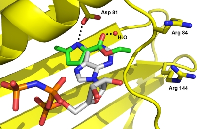
Fig 1.
Computational docking of a pyrrole NMR fragment hit overlaid with ATP in the crystal structure of the S. aureus GyrB 24-kDa N-terminal domain. Hydrogen bonds between the adenine of ATP, Asp81, and a water molecule (red sphere) are shown with dotted lines. Carbons of the pyrrole NMR hit are colored green, while those of ATP are colored gray. Other atoms are shown in standard coloring. The GyrB protein is colored yellow and displayed as a ribbon structure, with the exception of key amino acid side chains labeled by sequence number.

