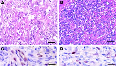
Figure 1.
Histologic patterns of cutaneous Kaposi sarcoma (KS) associated with a human herpesvirus 8 (HHV-8) type E infection. Patient 1: A) The spindle cells were organized as bundles, forming vascular slit-like spaces containing erythrocytes. Some macrophages containing hemosiderin were observed (data not shown). Scale bar = 25 μm. C) Immunohistochemical testing showed a positive signal for HHV-8 infection (latent nuclear antigen [LANA-1]) and CD34 (data not shown). The Perls staining also gave highly positive results (data not shown). Scale bar = 50 μm. (Patient 1 corresponds to the first patient [04/0480] in the Table A1], a 51-year-old mestizo man who had HIV-1 infection.) Patient 2: B) Spindle cells forming rare vascular channels, with numerous lymphocytes, plasma cells, and macrophages. Scale bar = 25 μm. D) Immunohistochemistry showed a lower positive signal for HHV-8 infection (LANA-1) and CD34 (data not shown). Few cells displayed a positive Perls staining (data not shown). Scale bar = 50 μm. (Patient 2 corresponds to the tenth patient [06/0772] in the online Table A1, a 24-year-old mestizo man with HIV-1 infection).

