Fig 2.
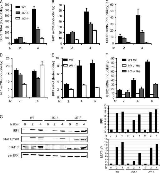
Regulation of IFN-γ-induced genes by IRFs. (A to D) WT, Irf3−/−, and Irf7−/− MEFs were treated with IFN-γ for the indicated time periods, followed by determination of GBP2, TAP1, SOCS1, and IRF1 mRNA expression by qPCR. (E) WT and Irf1−/− MEFs were treated with IFN-γ for the indicated time periods, followed by determination of IRF7 mRNA expression by qPCR. (F) WT, Irf1−/− and Irf7−/− bone marrow-derived macrophages (Mθ) were treated with IFN-γ for the indicated time periods, followed by determination of IRF7 mRNA expression by qPCR. GBP2, TAP1, SOCS1, IRF1, and IRF7 mRNA expression was determined by qPCR and normalized to GAPDH levels. (G) IRF1 protein expression and STAT1 tyrosine phosphorylation were detected by Western blot analysis. Differences in STAT1 expression levels between WT MEFs and MEFs deficient for IRF3 or IRF7 were analyzed by reprobing the blot with an antibody against the STAT1 C terminus. The Western blot was quantified by densitometry of the antibody-mediated signal (panel G, right), normalizing IRF1 and pYSTAT1 to the pan-ERK signal. qPCR measurements were made in triplicate. All experiments were repeated at least three times.

