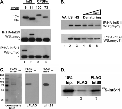
Fig 2.
Integrator 9 and Integrator 11 form a specific and robust heterodimer. (A) Upper panel, Western blot (WB) analysis of cell lysates from HeLa cells transfected with myc-tagged IntS9, IntS11, CPSF100, or CPSF73; middle panel, Western blot analysis of immunoprecipitates (IP) by the use of anti-HA agarose beads from cells transfected with HA-tagged IntS9 along with each of the myc-tagged proteins shown in the upper panel; lower panel, same as the middle panel except that data represent cells cotransfected with HA-tagged IntS11. The asterisks represent the cross-reacting Ig heavy chain present after immunoprecipitation. (B) Western blot analysis of cells cotransfected with HA-IntS11 and myc-IntS9 or with HA-IntS9 and myc-IntS11 (lower panel). Lysates were subjected to immunoprecipitation with anti-HA antibodies under various lysis and wash conditions (LS, 150 mM NaCl; HS, 500 mM NaCl; Denaturing, 0.1% SDS and 0.5% SDC) (lane 4) and then supplemented with either 500 mM NaCl (lane 5) or 1 M NaCl (lane 6). “VA” denotes lysates transfected with myc-tagged proteins with empty HA vector. (C) SDS-PAGE analysis of eluted FLAG-tagged, full-length IntS9 by the use of Coomassie blue staining (left panel) or Western blot analysis with FLAG antibodies (middle panel) or IntS9 antibodies (right panel). “—,” lane loaded with elution buffer only. (D) Results from a pulldown assay using FLAG-tagged IntS9 and [35S]methionine-labeled IntS11. The first lane represents 10% of the input (Inp.) IntS11, the middle lane reflects the amount of IntS11 pulled down with anti-FLAG beads alone, and the last lane represents the pulldown of IntS11 with IntS9.

