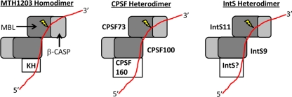
Fig 7.
Model of IntS9/11 binding to the 3′ end of snRNA compared to the known archaeal homodimeric CPSF-KH protein, representing the molecular model proposed by Silva et al. (31) based on their crystal structure of the MTH1203 β-CASP protein. This model is compared with a model for both the CPSF73/100 and IntS9/11 heterodimers. The MBL domain is represented in dark gray, the β-CASP domain in light gray, and the RNA substrate in red; the lightning bolt indicates the cleavage site.

