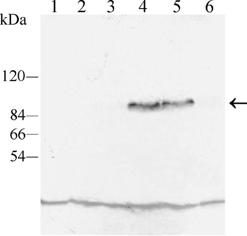
FIG. 8.
Coimmunoprecipitation of Tpk1-GFP with Bcy1p. S-100 fractions from strains CAI4, H2D, bcy1 tpk2, ASM1, ASM2, and ASM3 containing the same amounts of protein were subjected to immunoprecipitation with anti-C. albicans Bcy1p antiserum as described in Materials and Methods. Immunoprecipitates were resolved in SDS-7.5% PAGE, transferred to polyvinylidene difluoride membranes, and developed with a monoclonal anti-GFP antibody. Lanes: 1, parental strain CAI4; 2, mutant H2D strain; 3, bcy1 tpk2 double mutant strain; 4, ASM1 strain; 5, ASM2 strain; 6, ASM3 strain. The positions of molecular mass markers are indicated on the left. The arrow indicates the position of the 96-kDa fused protein band. The low-molecular-mass band observed in all lanes corresponds to a nonspecific reaction of the antibody.

