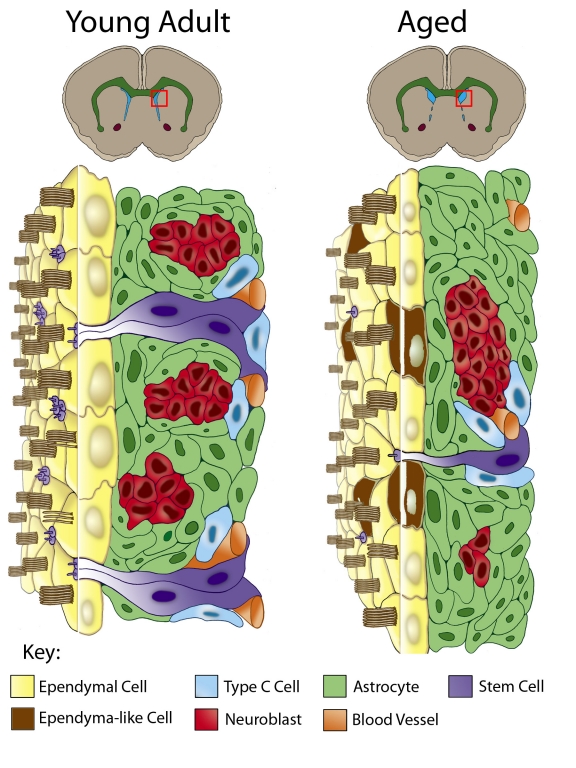
Figure 1.
Comparative cytoarchitecture of the SVZ through aging. Due to age-related stenosis of the ventral lateral ventricle walls, only the dorsolateral SVZ remains proliferative and the dorsal ventricle (blue in upper coronal brain images) expands. The red box in each schematic represents the dorsolateral SVZ depicted in more detail below. The young adult SVZ is organized below an ependyma monolayer (yellow cells) and includes astrocytes (green cells), astrocytes with an apical and basal process spanning the SVZ (NSCs, purple cells), neuroblasts (red cells), Type C cells (blue cells) and basally located blood vessels (orange). In the aged SVZ there is a significant reduction of the SVZ, with fewer astrocytes possessing an apical process and fewer neuroblasts and Type C cells. Additionally, some SVZ astrocytes (brown cells) are found incorporated within the ependymal monolayer. These integrated astrocytes are derived from dividing astrocytes and take on ependyma-like characteristics.

