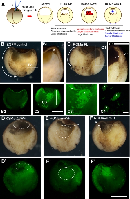
Fig 8.
Embryos overexpressing RGMa constructs exhibit abnormal internal morphology during gastrulation. (A) Schematic of bisected Xenopus gastrula embryos following overexpression of FL-RGMa, RGMa-ΔvWF, or RGMa-ΔRGD mRNA. Red descriptions indicate the RGMa-ΔvWF-specific phenotypes, whereas blue descriptions indicate the RGMa-ΔRGD-specific phenotypes. A, animal; V, vegetal. (B to F) Bisection of a control EGFP-injected embryo (B, B1, and B2) and RGMa mutant embryos (C to F, C1 to C3) that were collected when control embryos reached midgastrula. Panels B2 and C2 show fluorescent images of the same embryos shown in panels B and C, respectively. All bisected embryos show animal (top), vegetal (bottom), dorsal (left), and ventral (right) sides. (B and C) RGMa-overexpressed embryos show thicker ectoderm and reduced mesoderm involution. (B1 and C1) Magnified images of ectoderm from control and FL-RGMa-overexpressing embryos. Arrows in panels B, B1, C, and C1 indicate the different thicknesses of ectoderm and the different rates of mesoderm involution. (C3 and C4) Magnified images of C2 (C3) and isolated blastocoel cells (C4) showing aggregation of RGMa-positive cells (asterisks). These cell aggregates were not adherent to the blastocoel floor. (D and E) Overexpression of RGMa-ΔvWF generated two phenotypes. (F) An RGMa-ΔRGD mutant embryo exhibiting a phenotype similar to that for FL-RGMa-overexpressed embryos but showing a smaller blastocoel (dotted line). Facing arrows in panels D to F indicate the thickness of ectoderm adjacent to the tip of the involuting mesoderm. Bars, 300 μm (B, C, and D to F), 150 μm (B1 and C1), 300 μm (B2 and C2), and 150 μm (C3 and C4).

