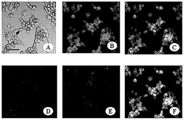
FIG. 2.

Confocal Immunofluorescence Microscopy demonstrating the binding of K2-3f to the mouse neuroblastoma N1E-115 cell line transfected with and stably expressing hDAT. Cells were fixed with 1% paraformaldehyde (A) and incubated with: 10 μg/ml K2-3f followed by PE -conjugated anti-mouse IgG (B), 20 μg/ml of polyclonal goat anti-hDAT IgG followed by FITC-conjugated anti-goat IgG (C), both (F), anti-OVA as a control (E). Untransfected N1E-115 cells as a background control were incubated with anti-hDAT antibody and then FITC-conjugated anti-goat IgG antibody (D). Confocal images were generated on an Olympus FluoView 300 confocal laser scanning system with an Olympus BX50 microscope. Bar, 50 μm.
