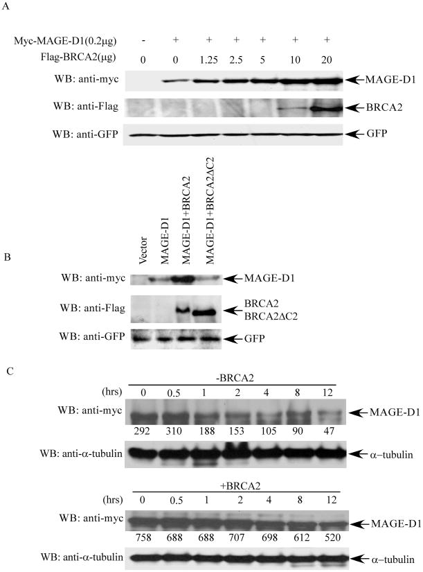
Fig. 3. BRCA2 expression stabilizes MAGE-D1.
(A). MAGE-D1 protein levels when co-transfected with increasing amounts of BRCA2. 293T cells were transfected with 0.2 μg pBB14-GFP and 0.2 μg Myc-tagged MAGE-D1 alone or with increasing amounts of Flag-tagged BRCA2, as indicated. Cell lysates were prepared 48 hr after transfection and analyzed for Myc-MAGE-D1 and Flag-BRCA2 protein levels by immunoblotting using anti-Myc or anti-Flag antibodies. Immunoblotting using anti-GFP antibody was used to control transfection efficiency. (B). MAGE-D1 protein level when co-transfected with wild-type BRCA2 or BRCA2δC2 (BRCA2 mutant devoid of the MAGE-D1 binding domain). (C). Half-life of MAGE-D1. 293T cells were transfected with Myc-tagged MAGE-D1 alone (upper panel) or with Flag-tagged BRCA2 (lower panel). Cycloheximide (final concentration 10 μg/ml) was added 48 hr after transfection to inhibit nascent protein synthesis. Cell lysates were prepared at the indicated time points after the addition of cycloheximide, followed by immunoblotting using anti-Myc and anti-α-tubulin antibodies to determine the turnover of MAGE-D1 protein. The numbers under the panels are quantification analyses of the signals as obtained by use of LabWorks software (UVP, Inc, Upland, CA).

