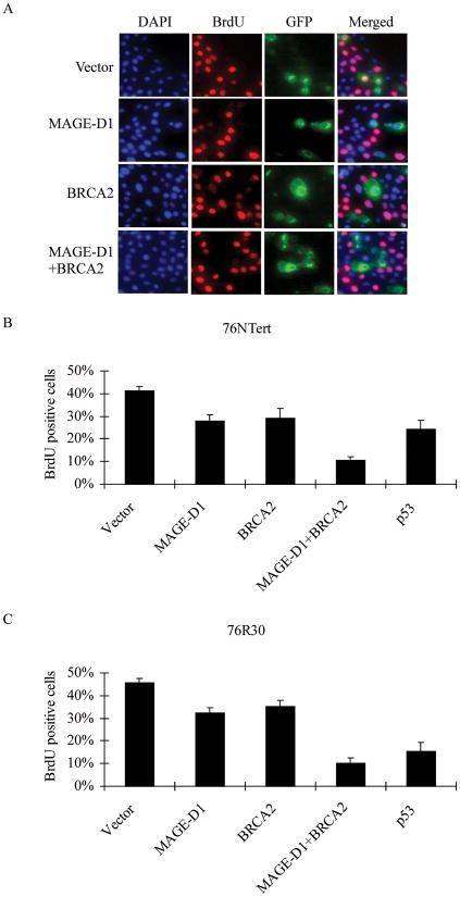
Fig. 4. BRCA2 and MAGE-D1 synergistically suppress cell proliferation.
(A) BrdU incorporation assay in 76NTert cells. 76NTert cells plated on coverslips in 24-wells plate were transfected with 0.5 μg MAGE-D1 and 1.5μg BRCA2, alone or in combination, together with pBB14-GFP (0.2 μg). The cells were incubated for 48 hr after transfection, then labeled with BrdU for 1 hr. The cells were fixed and stained using an anti-BrdU antibody. The DNA was stained with DAPI. Note that most of BRCA2 or MAGE-D1 transfected cells (GFP-positive cells) are negative for BrdU staining, while many vector- transfected cells (GFP-positive) or untransfected cells (GFP-negative) are positive for BrdU staining. (B and C) BRCA2 and MAGE-D1 synergistically suppress cell proliferation. 76NTert or 76R30 cells were transfected with MAGE-D1 and BRCA2, alone or in combination, as indicated, and assayed for BrdU incorporation as described in (A). The percentage of cells positive for BrdU staining was quantified in at least 300 transfected cells (GFP-positive cells). Each value represents the average and standard deviation from three independent experiments. The percentage of cells positive for BrdU staining was significantly lower in cells transfected with BRCA2, MAGE-D1, or MAGE-D1+BRCA2 than in cells transfected with vector alone (P < 0.05).

