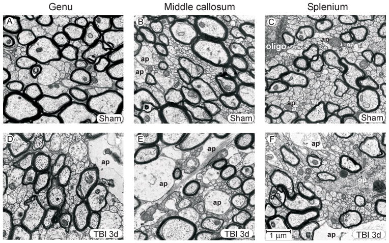
Figure 3.
Representative micrographs, showing genu, mid-callosum, and splenium regions in sham injured control rats (A–C) and during the post-injury period when fluid percussion injury-induced change in the unmyelinated axons was well developed (3 days post-injury) (D–F). Even though quantitative analyses revealed significant morphometric changes to axonal dimensions at 3 days post-injury, the general architecture of injured tissue was similar to that in sham control tissue, although in some injured rats astrocyte processes were larger and more frequently encountered. The rostral-to-caudal increase in unmyelinated axon density, noted in sham control rats, is preserved in the post-injury samples. Abbreviations: ap, astrocyte process; oligo, oligodendrocyte cell body; op, oligodendrocyte process. Calibration bar in F applies to all panels.

