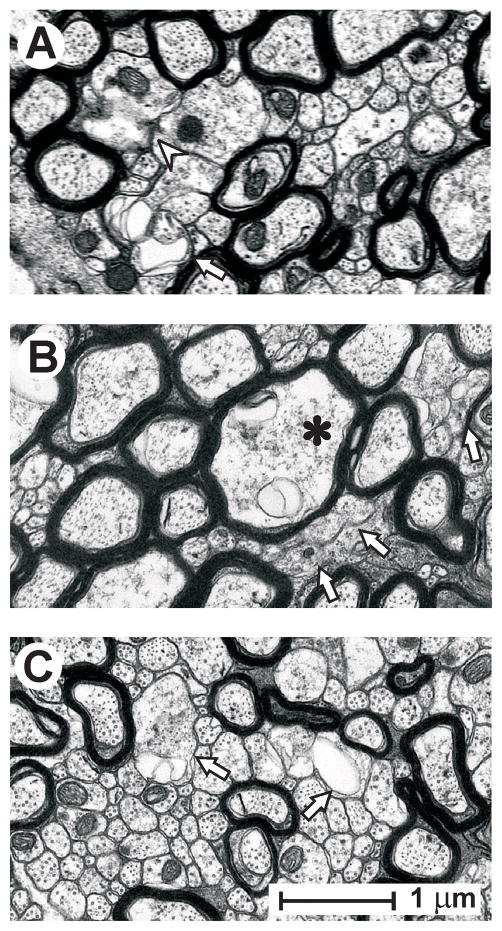
Figure 5.
Representative axonal profiles that failed to meet the operational definition of ‘intact’ in the splenium at 3 days post-injury. (A) Isolated unmyelinated axons exhibiting membrane discontinuities (arrowhead) and membranous folding apposed to aberrant extracellular spaces (arrow). (B) Clusters of unmyelinated fibers lacking distinct membranes (arrows), along with a myelinated axon (asterisk) with cytoplasmic abnormalities. (C) Example of vulnerability of relatively large unmyelinated axons (arrows) juxtaposed to intact small unmyelinated axons. All calibration as in panel C.

