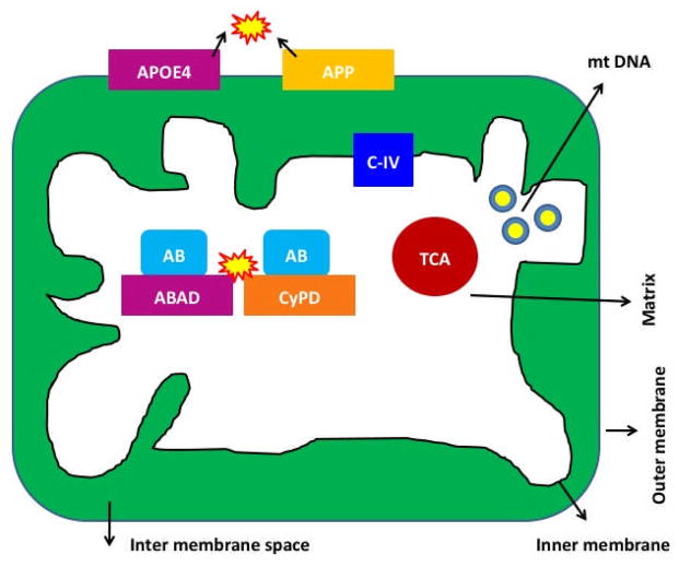
Figure 3. Localization of mutant proteins of AD in the mitochondria in neurons affected by AD.
In early-onset AD, genetic mutations of APP, PS1 and PS2 genes activate beta and gamma secretases, cleave from c-terminal region of amyloid beta precursor protein, and produce Aβ peptides. The toxic Aβ peptides enter mitochondria, interact with mitochondrial proteins, including cyclophilin D and ABAD --induce free radicals, decrease cytochrome oxidase activity, inhibit ATP generation and damage mitochondria both structurally and functionally. Further, in late-onset AD, APP is transported to outer mitochondrial membrane, blocks the import of nuclear cytochrome oxidase proteins to mitochondria and may be responsible for decreased cytochrome oxidase activity. In addition, N-terminal portion of ApoE4 is associated with mitochondria, induce free radicals and cause oxidative damage. In AD, complex IV of mitochondrial respiratory chain is affected.

