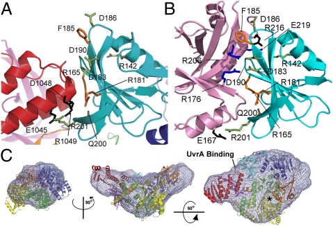
Fig. 2.
TRCF autoinhibition of UvrA binding is mediated by domain D7. (A) D2–D7 interaction seen in the crystal structure of apo TRCF (PDB ID 2EYQ). Conserved D7 residues are shown in black; interacting D2 residues are shown in green. Substitutions of orange residues are functionally important (21). (B) TRCF–UvrA interface. Residues in UvrA that bind UvrB are shown in blue (20). Other interacting residues are colored in black or as in A. (C) Crystal structure of apo E. coli TRCF (colored as in Fig. 1A) docked into the SAXS envelope. An asterisk indicates the ATP-binding site.

