Fig. 3.
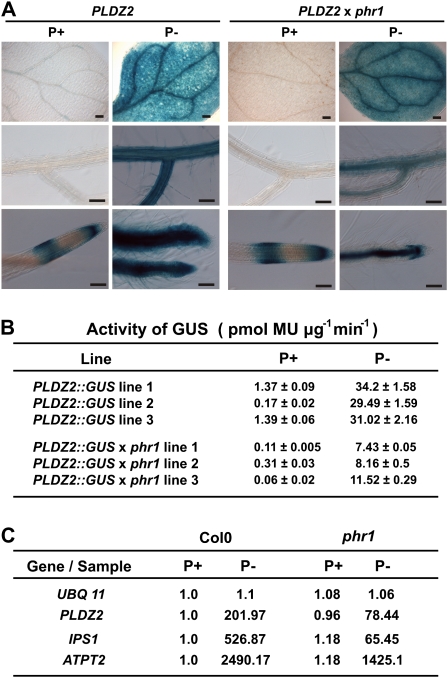
Expression of PLDZ2::GUS in Arabidopsis Col0 and phr1. (A) Tissue-specific expression of PLDZ2::GUS in 10-day-old seedlings grown for 10 days in media supplemented with 1 mM (P+) or 0 mM Pi (P–), subjected to histochemical GUS assays, and photographed using Nomarsky optics. (B) GUS specific activity of PLDZ2::GUS in Col0 and phr1 seedlings grown under P-sufficient and P-limited conditions. (C) Quantitative reverse-transcription PCR analysis of PLDZ2, IPS1, and ATPT2 in Col0 and phr1. Expression levels are reported as relative expression of the corresponding gene in Col0 grown under Pi-sufficient conditions. UBQ 11 was used as a non-responsive control. (This figure is available in colour at JXB online.)

