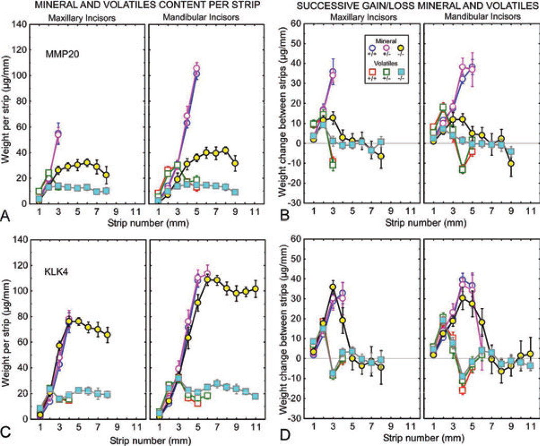
Fig. 3.
Mineral and protein contents in developing enamel on maxillary and mandibular incisors of mice with genetically altered expression of matrix metalloproteinase 20 (MMP20) (A, B) and kallikrein-related peptidase 4 (KLK4) (C, D). Mineral (circles) and protein (squares) weights per 1-mm-long enamel strip (A, C) and change in weights of both between sequential strips (B, D) for developing enamel on incisors from wild-type (Mmp20+/+, Klk4+/+; blue and red symbols), heterozygous (Mmp20+/−, Klk4+/−; mauve and green symbols), and null (Mmp20−/−, Klk4−/−; yellow and cyan symbols) mice. Each graph represents mean ± 95% CI. See Fig. 2A for the location of the stages of amelogenesis relative to each graph. The enamel from the mandibular incisors of Mmp20−/− and Klk4−/− mice is soft enough to allow strips to be cut across the length of the whole tooth (A–D). The enamel in Mmp20−/− mice is thinner than normal and disorganized (A, B), whereas the enamel in Klk4−/− mice is normal in thickness but softer than normal in depth (C, D).

