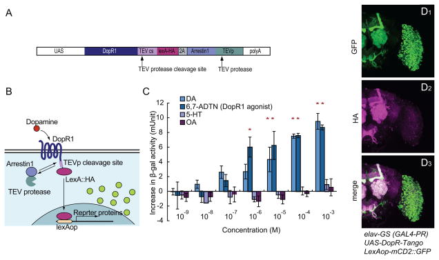
Figure 1. Characterization of DopR-Tango in vitro and in Drosophila.
(A) Design of the DopR-Tango transgene; note HA epitope tag on LexA.
(B) Schematic illustrating DopR-Tango mechanism.
(C) DopR-Tango reporter (β-gal) activity in response to indicated ligands in HEK293 cells co-transfected with CMV-GAL4, UAS-DopR-Tango and LexAop-β-gal. Increases in β-gal activity relative to background are shown. Error bars represent the standard error of mean (SEM). Asterisks represent statistically significant increases (p<0.05, t-test with Bonferroni correction, n=3).
(D) Representative confocal projections of whole-mount brains from DopR-Tango flies visualized with GFP native fluorescence (green) and anti-HA immunostaining (magenta).

