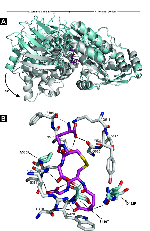
Figure 5.
Structure of HbpA2 from H. parasuis. (A) Ribbon diagram showing an overlay of GSSG-complexed GbpA (PDB id. 3M8U; gray) with HbpA2 from H. parasuis (PDB id. 3TPA; blue). The structures were superposed with respect to their C-terminal domains. HbpA2 shows a conformation that opens the cleft between the N- and C-terminal domains about 30° relative to its ligand-complexed paralogous counterpart. GSSG is depicted in atom-colored sticks. (B) Key binding residues of the GbpA C-terminal domain to accommodate GSSG (shown in atom-colored gray sticks) are replaced in HbpA2 by counterparts (shown in atom-colored blue sticks) that are incompatible with binding peptide-like allocrites. Residue numbering is according to PDB id. 3M8U. Some key interactions are depicted as black dashed lines. For clarity some interactions have been omitted. The figure was created with PyMOL (The PyMOL Molecular Graphics System, Schrödinger, LLC).

