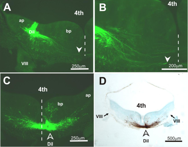
Figure 5.
Preliminary experiments to track the course of labeled fibers following DiI injection into the indicated sites in the hindbrain (E13.5). (A,B) Transverse section of the hindbrain at the level of the VIIIth cranial nerve root (VIII) showing DiI applied at a site approximately 150 μm into the alar plate and labeled projections coursing in a ventromedial direction towards the midline. Images were as viewed under epifluorescence microscopy with use of an FITC filter. Panel (B) is a magnified view of (A), showing growth cone-like terminals (solid arrowhead) of projection fibers approaching the midline. Dashed line denotes the midline. (C,D) Transverse section as in (A) but showing DiI applied into the floor plate (open arrowhead) for retrograde labeling of neurons. Images were as viewed under epifluorescence microscopy with use of an FITC filter (C), or after photoconversion of DiI labeling into a stable diaminobenzidine reaction product (D). The latter was counterstained with cresyl violet. Most cells that were so labeled are identifiable within the prospective area of the reticular formation; few, if any, labeled cells are observable in the area of the VN. Abbreviations: 4th, fourth ventricle; ap, alar plate; bp, basal plate; cp, cerebellar primordium; V, Vth cranial nerve root; VIII, VIIIth cranial nerve root; r2 to r7, rhombomeres 2 to 7.

