Figure 8.
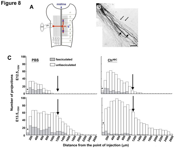
Quantification of DiI-tracked commissural projections of the VN. (A) Schematic diagram showing overlay of longituinal gridlines against DiI-tracked fiber projections (red) of the hindbrain flat-mounts. The injection site was set as '0' and projections to the right (contralateral) were counted. (B) Image of fasciculated fibers (thick arrows) mixed with unfasciculated fibers (thin arrows). (C) Histograms of fasciculated and unfasciculated projections against the distance from the DiI injection site in hindbrains treated with PBS (left panels) and with ChABC (right panels). Data are representative of five individual sets of experiments at E12.5(+1 DIV) and E13.5(+1 DIV). The downward pointing arrow indicates the position of the midline. Asterisks denote regions within 100 μm of the DI injection site where the intensity of dye precluded accuracy of fibre counts.

