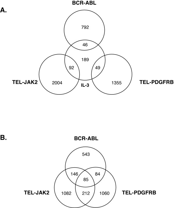
Figure 1.
Comparison of gene expression profiles among BCR-ABL, TEL-PDGFRB, TEL-JAK2 and IL-3. Ba/F3 cells were washed and incubated in IL-3-depleted media for 5 h, then stimulated with IL-3 for 0 h or 1 week. Ba/F3 BCR-ABL cells and Ba/F3 TEL-PDGFRB cells washed and incubated in the media depleted of IL-3 and supplemented with Imatinib for 5 h, were washed and incubated in the absence of IL-3 and Imatinib for 0 h or 1 week. Ba/F3 TEL-JAK2 cells were washed and incubated in IL-3-depleted media for 1 week. Total RNA was collected at 0 h and 1 week time-points, and was used for microarray analysis. For all cell lines except for Ba/F3 TEL-JAK2 cells, changes in gene expression were calculated using the expression values at 1 week and the 0 h within each cell type. For Ba/F3 TEL-JAK2 cells, expression values obtained from Ba/F3 TEL-JAK2 cells at the 1 week time-point were compared to the expression values obtained from Ba/F3 cells at the 0 h time-point. The number of genes that were induced or suppressed by 2-fold or more with significant detection scores in each cell type as well as in overlapped regions is indicated in the Venn diagrams.

