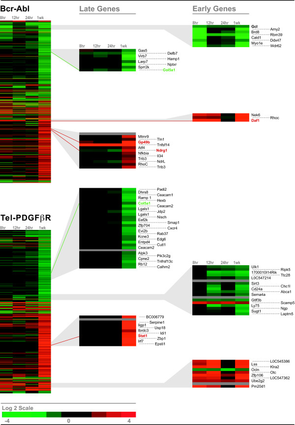
Figure 2.
Comparative gene expression profiling between genes regulated by BCR-ABL and genes regulated by TEL-PDGFRB. Ba/F3 BCR-ABL cells and Ba/F3 TEL-PDGFRB cells were washed and incubated in media depleted of IL-3 and supplemented with Imatinib for 5 h. Cells were washed and incubated in the absence of IL-3 and Imatinib to activate the fusion kinases for 0, 8, 12, 24 h and 1 week. Total RNA was collected at each time-point and was used for microarray analysis. The 250 most highly induced and the 250 most highly suppressed genes at 1 week were identified in cells with BCR-ABL (Top) or TEL-PDGFRB (Bottom) fusion proteins. The 500 genes induced or suppressed by each fusion protein were hierarchically clustered using the Eisen Cluster and Tree View programs and are displayed on a log2 color scale, with red representing induced genes and green representing suppressed genes. Grey represents omitted data. Shown in the second and the third columns are the examples of genes that are regulated by 2-fold or greater within 24 h of activation ("Early Genes"), and after 24 h ("Late Genes"). Genes that were validated by Q-PCR are highlighted in red (induced) or green (suppressed) in the gene list.

