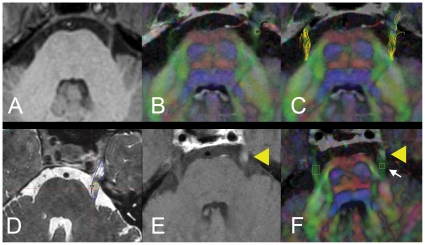
Figure 1. Baseline MR imaging, tractography of the trigeminal nerve, target and ROI definition.
Image processing commenced with baseline anatomical 3TMR images (A, axial section, midpontine level). Diffusion tensor images with overlaid colour-by-orientation fibers are shown in B. Reconstructed tracts of the trigeminal nerve onto colour-by-orientation images are shown in C. Panel D depicts the contour of the trigeminal nerve (blue) and location of the radiosurgical shot. Yellow circle denotes the 80% isodose line, representing the “target” of Gamma radiation to the nerve. Panel E shows focal area of post-gadolinium enhancement on the trigeminal nerve (yellow arrowhead), defining the “target” ROI. Panel F shows the location of the “proximal” ROI, proximal to the area of gadolinium enhancement (B, white arrow), and “unaffected” ROI, contralateral nerve.

