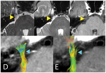
Figure 5. Tractography can detect changes in the trigeminal nerve in the absence of post-treatment gadolinium enhancement: Panels A to E delineate FA changes seen after treatment. Subject S2 did not show post-treatment MR gadolinium enhancement.
Panel A shows location of radiosurgical target during treatment planning. Panels B, C depict post-treatment MR and lack of gadolinium-enhancement (yellow arrowhead). Reconstructed trigeminal tracts are shown in panel D (pre-treatment) and E (post-treatment), with clear FA changes in the target area (blue arrowhead).

