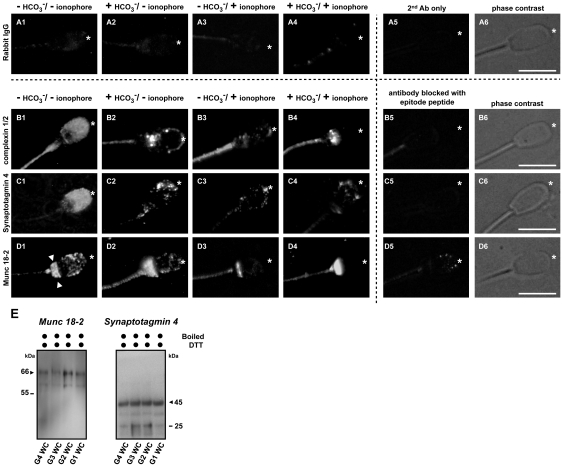
Figure 3. Bicarbonate-dependent relocalization of SNARE interacting proteins.
(A–D) Immunofluorescence detection of SNARE interacting proteins complexin 1/2, synaptotagmin 4 and Munc 18-2 showed a homogenous distribution over the entire sperm head in the control, non-activated sperm (B1–D1). Munc 18-2 had an additional strong staining at the post-equatorial region (D1, indicated with arrow heads). Upon capacitation, these proteins relocalized to the apical sperm head (marked with asterisk) and appeared as punctate aggregates indicating their association with other proteins (B2–D2). When AE was induced (either in the absence [B3–D3] or in the presence [B4–D4] of bicarbonate), the majority of the signal at the apical ridge of the sperm head disappeared, which was due to the removal of apical membranes after shedding MVs from the sperm heads. A minor signal can still be detected in some sperm cells when AE is induced in the absence of bicarbonate while the majority of the signal was lost in the acrosome reacted sperm in the presence of bicarbonate; this is due to different efficiencies in AE induction. Purified rabbit IgG were used as a negative control (A1–A4) since these Abs were raised in rabbit. To demonstrate the specificity of the signals observed, sperm cells were also incubated in the absence of primary Ab (A5) or were incubated in the primary Ab that was immunized with specific blocking peptide of which the antibody was produced (B5–D5). Bars represent 10 µm. (E) Validation the presence of Munc18-2 and synaptotagmin 4 in porcine sperm. Whole sperm cell lysates (WC) from 4 different incubation conditions (G1–G4, see Methods and Materials) were used for the further validation on the presence of Munc 18-2 and synaptotagmin 4 observed in other independent experiments. The arrows indicate the expected molecular weight of protein on the Western blot.

