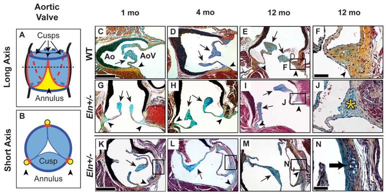
Fig. 1. Eln+/− aortic valves demonstrate cartilage-like nodules at all stages and annulus-specific proteoglycan accumulation at the aged stage.
Long (A, C–J) and short (B, K–N) axis sections of WT (C–F) and Eln+/− (G–N) aortic valves. The dotted line in A represents the plane of dissection shown in B. Pentachrome staining identified proteoglycans as blue in the cusp (arrows) and collagens as yellow in the annulus (arrowheads); this color coding is maintained in A and B with red lines representing the hinge. In aged (12 mo) Eln+/− valves, proteoglycan accumulation was observed in the annulus region (asterisk, J). Cartilage-like nodules were present in the Eln+/− annulus at all stages (black boxes, K–M; arrow N). AoV, aortic valve; Ao, aorta. Scale bars: 20 μm (F, J, N); 120 μm (all others).

