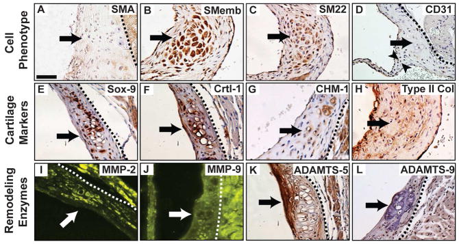
Fig. 2. Characterization of cartilage-like nodules in the Eln+/− aortic valve annulus.
The nodules (arrows) were localized in the annulus region and characterized by cell phenotype (AD), cartilage markers (E–H), and remodeling enzymes (I–L). CD31 positive cells are present in the interstitium (arrowheads, D). All sections are in short axis; derived from aged mice, except panels D and G (adult). Scale bar: 20 μm. Dotted lines isolate valve tissue from aortic and/or myocardial tissue.

