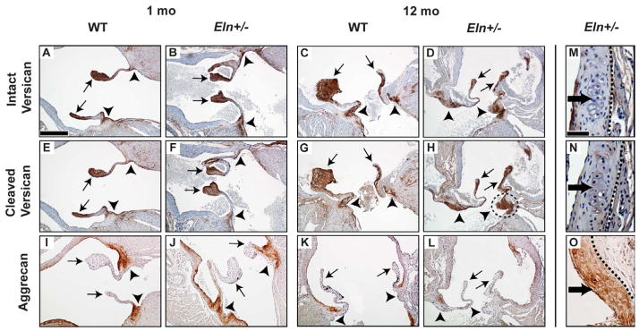
Fig. 3. Abnormal regional versican processing in Eln+/− aortic valves.
Increased cleaved versican was shown in the aged (12 mo) Eln+/− annulus region (dotted circle, H) compared to age matched WT (G), consistent with proximate ADAMTS-5 expression (Fig. 2K). Neither intact nor cleaved versican was expressed in the nodules (dark arrows, M–N), but aggrecan was (O). Cusps (arrows), annulus (arrowheads). Sections are oriented in long (A–L) or short (M–O) axis as illustrated in Fig. 1. Scale bars: 60 μm (A–L); 20 μm (M–O). For short axis sections, stage is 1 mo for M and N, and 12 mo for O. Dotted lines isolate valve tissue from aortic and/or myocardial tissue.

