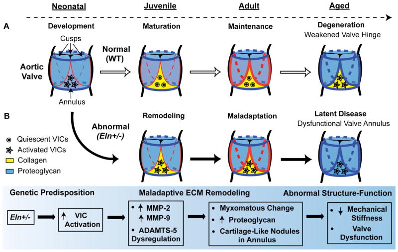
Fig. 8. Working model of regional structure-function relationships in latent AVD.
Color coding is based on the pattern of pentachrome stain with proteoglycans as blue and collagens as yellow. Normal valves (A, white arrows) undergo development, maturation, maintenance and age-associated degeneration over time (dotted arrow). The valve annulus (yellow) and hinge (red) mature during early postnatal growth. Abnormal valves (B, black arrows) are characterized by a genetic predisposition to AVM (Eln+/−), early maladaptive ECM remodeling mediated in part by VIC activation, cell-matrix abnormalities disproportionately affecting the annulus region, and ultimately abnormal structure-function due to biomechanical failure at the hinge and valve dysfunction. We speculate that pathogenesis is initiated in the valve annulus region resulting in maladaptive ECM remodeling, and overt disease manifests later due to structural failure at the valve hinge.

