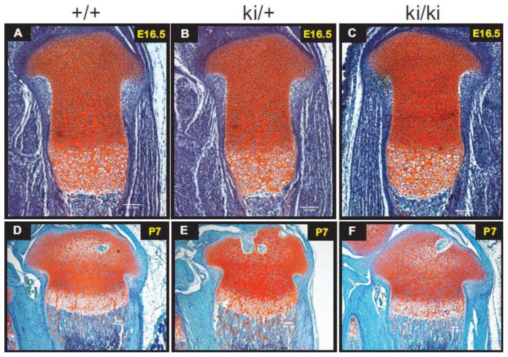
Fig. 4.
Safranin-O staining of wild type (+/+), heterozygous (ki/+) and homozygous knock-in (ki/ki) hindlimb tissue sections. Positive Safranin-O staining (red) is shown throughout the cartilaginous region of the developing growth plate and trabecular bone in E16.5 developing tibiae (Panels A–C) and in P7 developing proximal tibiae (D–F). Scale bars = 100 μm.

