Fig. 6.
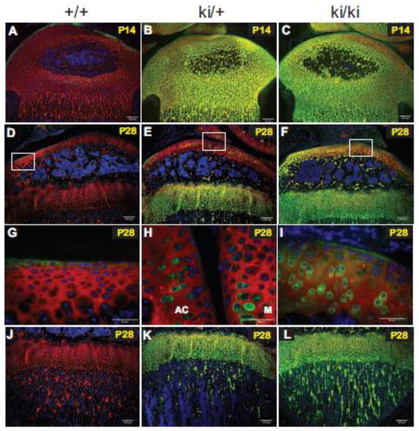
Type II procollagen protein expression patterns in P14 and P28 hindlimbs. Dual fluorescence immunohistochemistry was carried out to detect type II procollagen proteins in wild type (+/+), heterozygous (ki/+) and homozygous (ki/ki) proximal tibiae. Green staining represents anti-IIA positive proteins (i.e. the exon 2-encoded CR domain within the NH2-propeptide of type II procollagen or the α3 chain of type XI procollagen). Red fluorescent staining denotes the type II collagen triple helical domain. Areas of yellow fluorescence represent co-localization. Panels A-C, P14 tissue; Panels D-L, P28 tissue. Panels G, H and I are higher magnification images of the white boxed regions shown in Panels D, E and F, respectively. Note that the magnified image in Panel H has been rotated 90 degrees clockwise. AC=articular cartilage; M=meniscus (Panel H). Panels J–L show the metaphyseal trabecular bone region of developing tibiae. Scale bars = 100 μm (Panels A–F; J–L) or 50 μm (Panels G–I).

