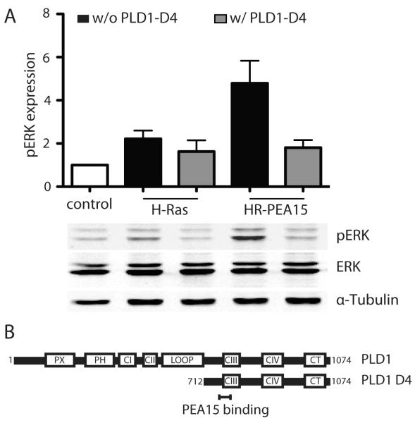
Figure 7.
Binding of PEA-15 to PLD1 is required for PEA-15’s effect on H-Ras mediated enhancement of ERK activity. (A) Western blot analysis of pERK and total ERK in iBMK cells transiently expressing combinations of H-Ras, PEA-15 and the PEA-15 binding domain mutant of PLD1 (PLD1-D4). Cells were kept in suspension for 18h. pERK levels are normalized to the total ERK expression. Shown is the mean and SEM of three independent experiments. n.s. not statistically significant (B) Structure of PLD1 and its PEA-15 binding site containing D4 domain used in the experiment. Boxes show functional domains, the PEA-15 binding site is highlighted. PX: phox homology domain, PH: pleckstrin homology domain, CI-CIV: conserved regions, CT: C-terminus.

