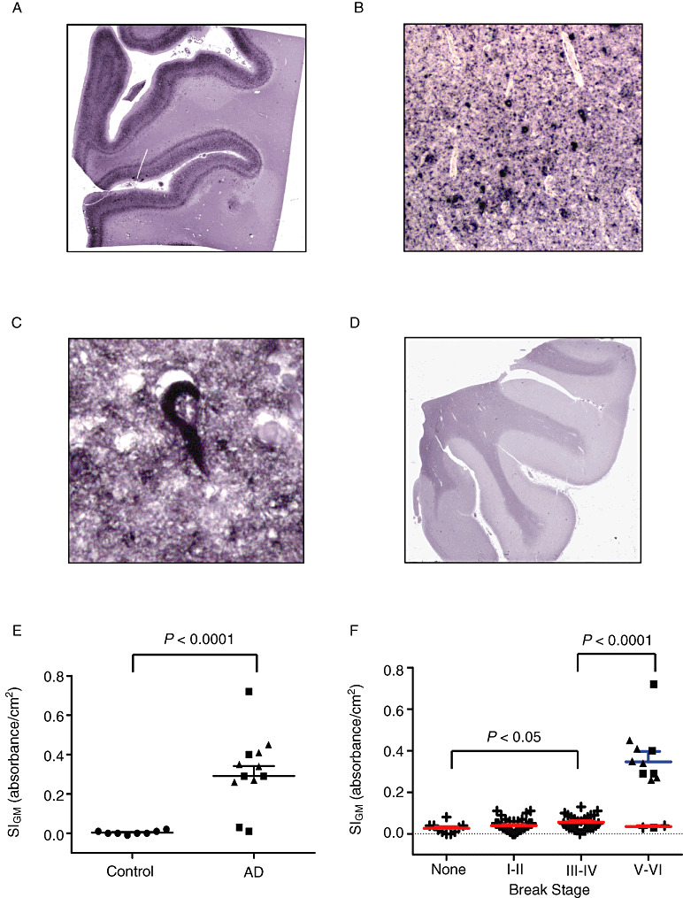
Figure 4.

Paired helical filament– (PHF‐) tau Histelide.
A. Section of middle frontal gyrus (MFG) from a late‐onset Alzheimer's disease (AD) (LOAD) case using Histelide technique. 5‐Bromo‐4‐chloro‐3‐indolyl phosphate (BCIP) deposits reveal the typical “ribbon” of superficial and deep PHF‐tau accumulation in cortical gray matter. B. Light microscopy showed abundant neurofibrillary tangles (NFTs) and neuropil threads (×100). C. Higher magnification (×400) revealed a classic NFT. D. Section of MFG from a control case; BCIP deposits are absent from cortical gray matter. E–F. Scatterplots for PHF‐tau signal intensity for gray matter (SIGM) in sections of MFG from control (●), late‐onset AD (■), autosomal dominant AD (▴), and Adult Changes in Thought (ACT) high‐cognitive performer (+) cases. Average (—) and standard error of the mean ( ) are indicated. E. Average PHF‐tau SIGM was significantly greater in both LOAD and autosomal dominant AD cases (n = 12) compared with controls (n = 8, P < 0.0001). F. PHF‐tau SIGM increased significantly with increasing Braak NFT stage among the ACT high performers (n = 79, P < 0.05), and was significantly greater in AD cases (n = 12) compared with ACT cases with Braak NFT stage III or IV (P < 0.0001).
