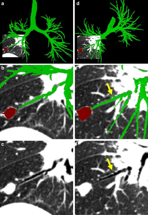
Fig. 3.
Illustration of system GUI interaction and route inspection (case 24). a The global 3D airway tree (green) fused with the local oblique cross-section view. The ROI, a suspect cancer nodule situated in the right lower lobe, appears in red. The oblique cross-section shown here and in other figures has isotropic pixel resolution 0.5 mm and uses display window [−1,000 HU, −200 HU]. b A close-up of the oblique cross-section near the ROI. c The same oblique cross-section as (b) displayed without the airway tree or ROI. d Another fused view after rotating the 3D scene to give another view of the small peripheral airway of (a–c). This airway is marked by yellow arrows in (e) and (f)

