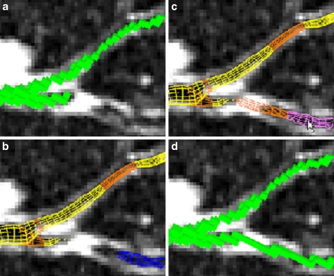
Fig. 4.
Example of airway tree augmentation for case 25 (scan dimensions, 512 × 512 × 796; voxel dimensions, ∆x = ∆y = 0.86 mm, ∆z = 0.50 mm; MDCT scanner, Siemens Sensation-16). a Current voxel-based local oblique cross-section view of the defined airway tree Tf before defect correction. b Wire frame surface representation of branch segments (yellow) and branch segment connections (orange) belonging to Tf and a branch segment (blue) not currently constituting part of the defined tree Tf. This view clearly indicates a missing branch defect. The disconnected branch segment Si ∉ S disconnects from the main defined airway tree because of a layer of high-intensity voxels and appears to have no visible child branches. c By selecting the missed branch segment, the user enables the segment Si and its previously computed and stored parent connection (i,P(i)) to be added to the defined tree Tf. d Voxel form of the resulting augmented airway tree segmentation

