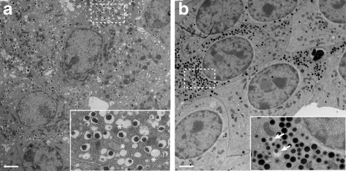
Fig. 1.
Appearance of ISGs in chemically and HPF fixed beta cells. Representative electron micrographs of beta cells in isolated pancreatic islets fixed either chemically (a) or with HPF (b); scale bars on the bottom left: 4 μm. The insets show a higher magnification (×3 the original) of the areas marked with a dashed rectangle. In (a), asterisks identify ‘ghost’ ISGs. In (b), arrows point to ISGs with halos

