
Figure 1.
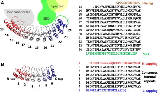
Ribbon diagrams and amino-acid sequences of D34 and NI6C. (A) The ribbon diagram of the D34 fragment of the membrane-binding domain of ankyrin-R (R13-R24, PDB 1N11 (11)). The beginning fragment of spectrin-binding domain (SBD) interacts with repeats R20–R24. Several ARs of ankyrin-R interact with the cytoplasmic domain of ion transporter, and SBD interacts with spectrin. The amino-acid sequence is shown in the right panel. (B) The ribbon diagram of NI6C. NI6C is composed of the N-capping repeat, the C-capping repeat, and six internal consensus repeats. (Right panel) The amino-acid sequence.