Figure 5.
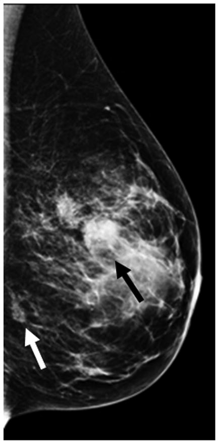
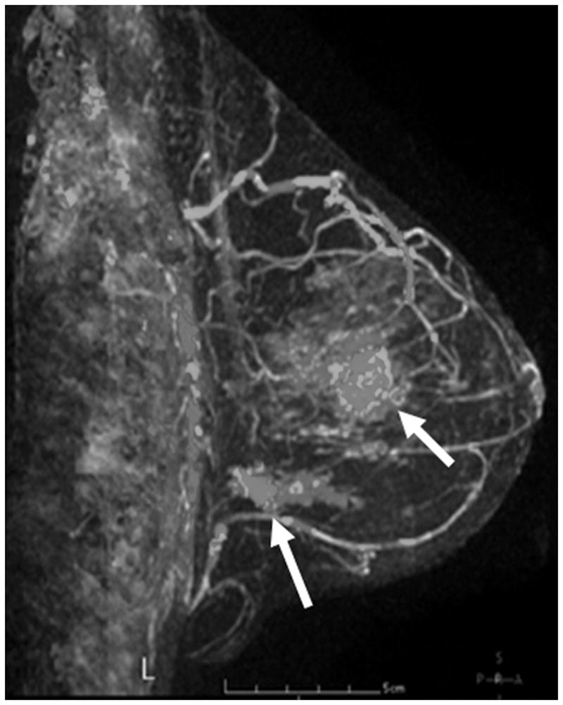
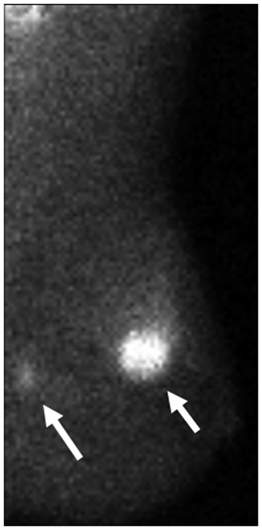
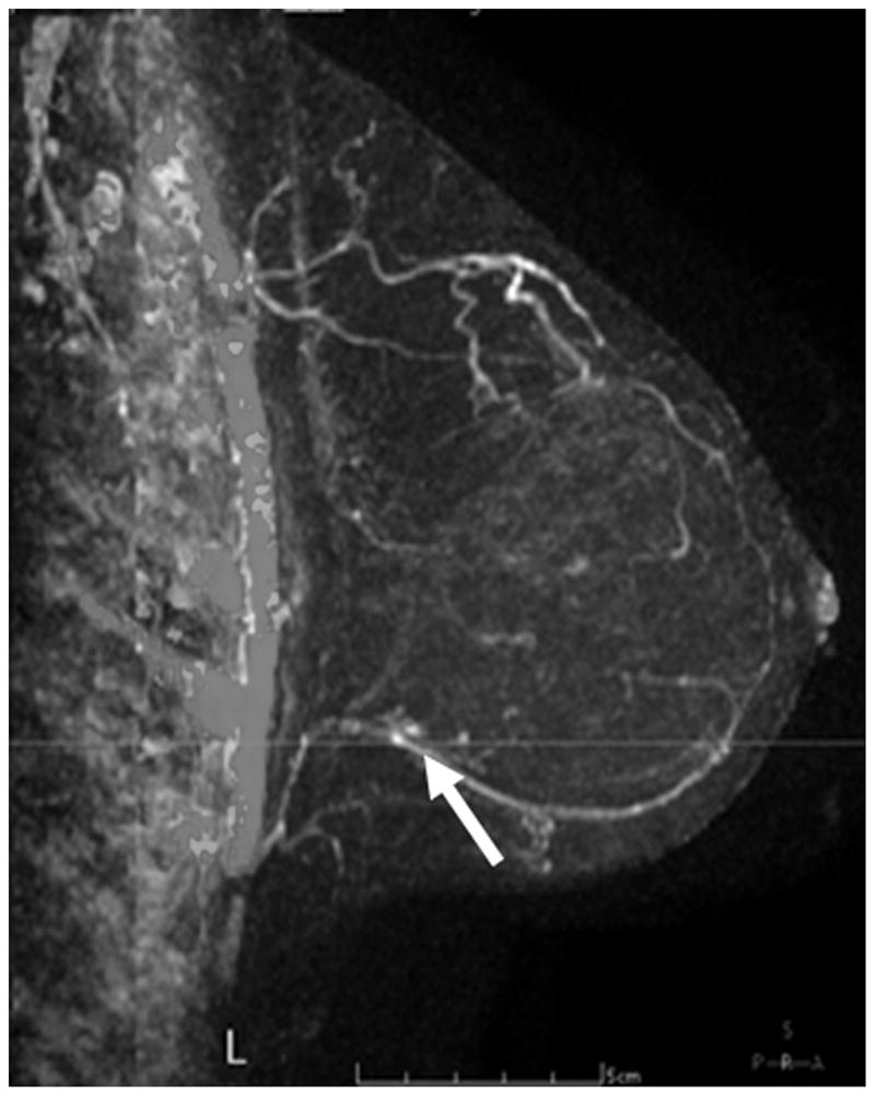
Pre–Neoadjuvant Therapy Imaging vs Post–Neoadjuvant Therapy Imaging. Pre–neoadjuvant therapy (pre-NT) imaging shows multifocal disease (A–D, arrows) by: (A) mammography (MMG), (B) magnetic resonance imaging (MRI), and (C) molecular breast imaging (MBI). Post–neoadjuvant therapy (post-NT) imaging (D) by MRI shows minimal enhancement remaining in lower outer breast and (E) by MBI is negative.
