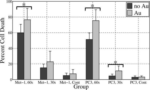
Fig. 7.
In vitro cell killing relative to 100% cell death control for cells not incubated with GNPs (dark gray) and cells incubated with 10 nm GNPs (light gray). Results from two cancer cell lines, Met-1 and PC-3 are shown. For each cell line, the treatment groups included 60 s and 30 s RF exposure at 100 W. The corresponding no RF controls (Cont) are also shown. With better than 95% confidence (p<0.05), the combination of GNPs and RF exposure demonstrated statistically significant greater cell death compared to the no GNPs for groups labeled with an asterisk (*). In all cases with GNPs, the cells were incubated with 3 μM 10 nm GNPs.

