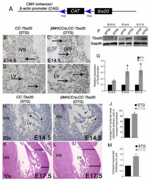
Figure 1. βMHCCre-mediated Tbx20 overexpression in differentiated cardiomyocytes results in thickening of ventricular myocardium.
(A) Schematic representation of the CAG-CAT-Tbx20 (CC-Tbx20) transgene for Cre-dependent Tbx20 expression. The CC-Tbx20 transgene consists of a CAG regulatory element (CMV enhancer linked to chicken β-actin promoter) that drives the ubiquitous expression of the CAT (chloramphenicol transferase) gene flanked by loxp sites. In the presence of Cre, the CAT gene and translational stop sequences are deleted and Tbx20 is expressed. (B-E) Overexpression of Tbx20 in E14.5 chamber myocardium of the βMHCCre;CC-Tbx20 double transgenic (DTG) animals is detected by Tbx20 antibody reactivity visualized by DAB staining. Increased Tbx20-positive dark brown/black nuclei (arrows) are apparent in the (C) interventricular septum (IVS) and (E) left ventricular (LV) myocardium of a DTG heart compared to a single transgenic (STG) littermate control (B,D). (F, G) Quantitative Western blot analysis reveals a 1.7-fold, 3.2-fold and 3.4-fold increase of Tbx20 protein expression in E12.5, E14.5 and E17.5 DTG hearts, respectively, compared to littermate STG controls. (H-J) In H&E stained sections, decreased trabecular myocardium is apparent in E14.5 DTG LV (compare asterisks in H versus I) and the average number of cells per area of the compact myocardium is increased compared to littermate control (J) (n = 4). (K-L) The thickness of the compact myocardium (black lines) is significantly increased in the E17.5 DTG heart as compared to a littermate control in H&E stained sections. (M) The compact layer thickness was quantified (n = 4) with statistical significance determined by Student’s t test, where * denotes p < 0.05.

