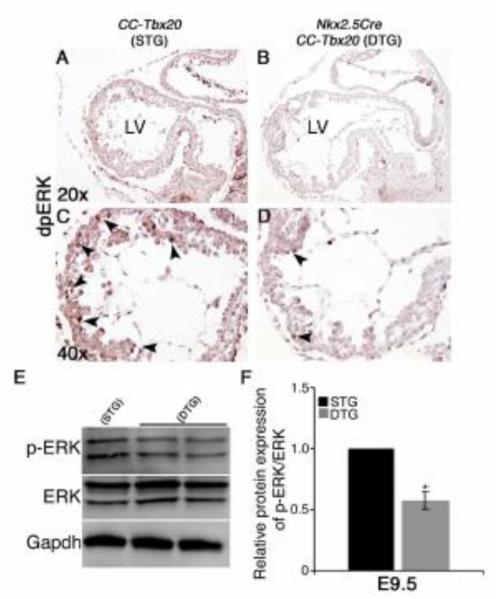
Figure 8. Nkx2.5Cre-mediated overexpression of Tbx20 results in decreased dpERK expression in embryonic cardiomyocytes.
(A-D) Expression of the diphosphorylated form of ERK1/ERK2 (dpERK) in E9.5 chamber myocardium is detected by dpERK-specific antibody reactivity visualized by DAB staining. Panels C and D represent higher magnified views (40x objective) of the LV region from panels A and B (20x objective), respectively. Reduced dpERK reactivity (brown/black nuclei) is apparent in the ventricular cardiomyocytes (arrowheads in D) of a DTG embryo compared to the STG littermate control (arrowheads in C). (E-F) dpERK expression was quantified by Western blot analysis as the ratio of dpERK to total ERK protein in E9.5 DTG hearts compared to littermate STG controls (n = 6-8). Statistical significance is determined by Student’s t test, where * denotes p < 0.05.

