
Figure 7.
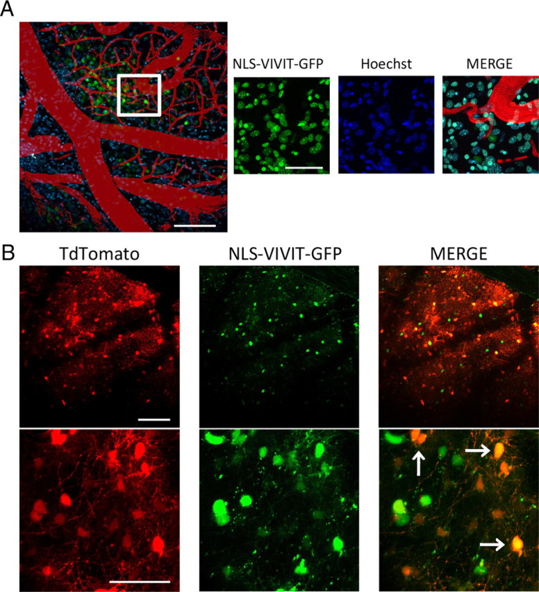
In vivo, NLS-VIVIT-GFP is targeted to the nucleus and coinjection of both AAV-NLS-VIVIT and AAV-Tdtomato efficiently transduces the same cells. A, Representative in vivo images of a mouse injected with AAV-NLS-VIVIT-GFP (left) confirm that the localization of NLS-VIVIT is restricted to the nucleus and colocalizes with Hoechst applied topically (right). Scale bars: 100 μm on the left and 50 μm on the right. B, Coinjection of AAV-NLS-VIVIT-GFP and AAV-Tdtomato with an appropriate ratio (3:1) is an efficient approach to be able to detect spines in vivo and to ascertain the fact that the observed Tomato-transduced cells also express NLS-VIVIT (arrows indicate GFP/Tdtomato double positive cells). Scale bars: 100 μm on the top and 50 μm on the bottom.