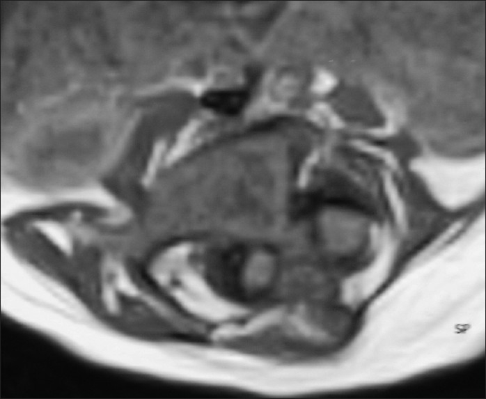
Figure 2.

Magnetic resonance imaging of the spine axial section showing evidence of diastematomyelia with a large intraspinal spur at L2–L3 vertebral level. The two hemicords were asymmetric; left one smaller than right and each hemicord was seen in a separate dural sac
