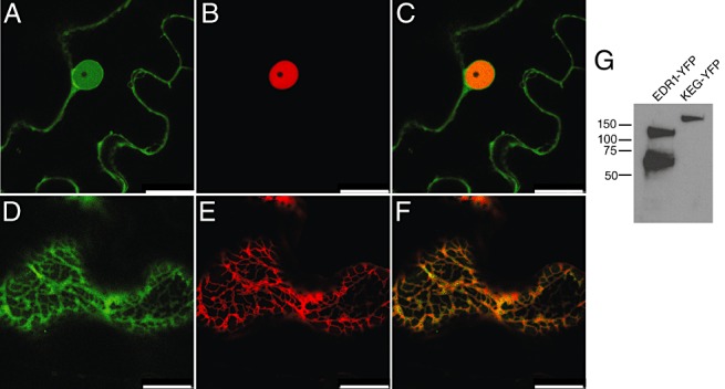
Figure 3.

Subcellular localization of enhanced disease resistance 1 protein (EDR1). (A–C) EDR1‐sYFP and GCN5‐mCherry were transiently co‐expressed in Nicotiana benthamiana leaves and imaged using confocal laser scanning microscopy. (A) EDR1‐sYFP (a single optical section taken through the nucleus of an epidermal cell). (B) GCN5‐mCherry expressed in the same cell. (C) Overlay of (A) and (B). (D–F) EDR1‐sYFP and mCherry ER marker (see Experimental procedures) were transiently co‐expressed in N. benthamiana. (D) EDR1‐sYFP (a single optical section taken through the cell cortex of an epidermal cell). (E) mCherry‐HDEL. (F) Overlay of (D) and (E). (G) Immunoblot of EDR1‐sYFP extracted from N. benthamiana leaves. KEG‐sYFP is an unrelated yellow fluorescent protein (YFP) fusion protein included to show the specificity of the antibody. Scale bar, 25 µm.
