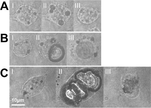
Fig. 2.

Bac-1 cells incubated with 90 nm gold spheres at 4.5∙109 part/ml and irradiated at 532 nm: (I) before irradiation, (II) with a delay equal to half the bubble lifetime after irradiation with a single laser pulse and after the trypan blue staining procedure. (A) Radiant exposure = 45 mJ/cm2, Δt = 300 ns. Absence of staining after application of trypan blue (III) suggests an intact cell membrane. (B) Radiant exposure = 80 mJ/cm2, Δt = 500 ns. The staining (III) indicates acute membrane damage. (C) Radiant exposure = 330 mJ/cm2, Δt = 190 ns. The cell is destroyed beyond recognition (III). Note that this cell was not stained. The staining procedure washes away the remnants of the cell. Scale bar: 10 μm, applicable to all images.
