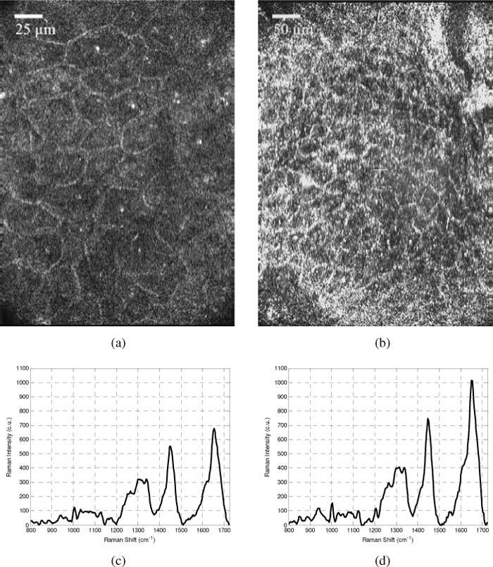
Fig. 6.

Images and spectra acquired from the fingertip using the 100x objective and hemisphere (a,c) and 50x objective and hemisphere (b,d). Cell membranes and nuclei are easily distinguished in (a), but nuclear detail is less evident when using the lower magnification objective (b). Image size is 260 x 198 µm and 520 x 396 µm for (a) and (b) respectively. Corresponding Raman spectra demonstrate the fact that the 50x objective allows for greater detected Raman signal intensity.
