
Fig. 2.
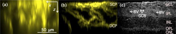
x-z (axial) images of fixed and stained mouse retina as acquired with conventional wide-field fluorescence (a), structured illumination fluorescence (b) and full-field optical coherence (c) microscopy techniques. Images (a)&(b) were constructed as the maximum value pixel projection from 512 x-z sections of the reconstructed images. Capillary plexi: SCP –superficial capillary plexus; DCP—deep capillary plexus; Retinal layers: GCL—Ganglion cell layer; IPL—Inner plexiform layer; INL—Inner nuclear layer; OPL—Outer plexiform layer; ONL—Outer nuclear layer; Arrows indicate: BV—Blood vessel; GCB—Ganglion cell bodies.